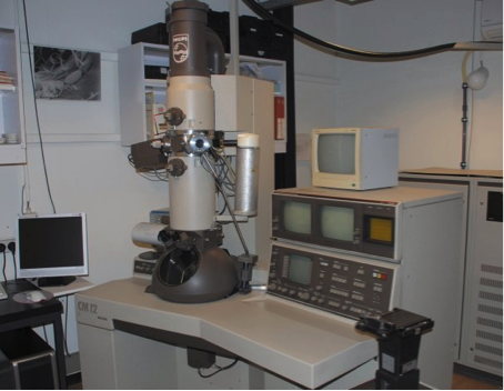
SCANNING AND TRANSMISSION ELECTRON MICROSCOPY
|
DESCRIPTION Acquisition in 1991, upgraded with a digital camera in 2004. Images in transmission mode (TEM) and Scanning mode (SEM). Equipped with the following detectors: Bright field, dark field, SED (Secondary Electron Detector) and BSED (Back Scattered Electron Detector). Imaging is done by the MegaView III digital camera in TEM and the slow scan device from the microscope in scanning mode. Both systems are piloted by Soft Imaging System from Olympus.
|
|
APPLICATIONS
|
Transmission electron microscopy with possibility of cytochemistry From : B. K. Kishore et al (1996),
|
Scanning electron microscopy From: Mettlen et al (2006),
|
Immunogold surface labelling by Platinum-Carbon Replicas From: Van Der Smissen et al (1992), |
COSTS
Cost : 50€ / h for the electron microscope
Costs for specimen preparations has to be evaluated and depends on type of treatment, colorations or coatings
Contact : Donatienne Tyteca or Patrick Van der Smissen


