APPLICATIONS
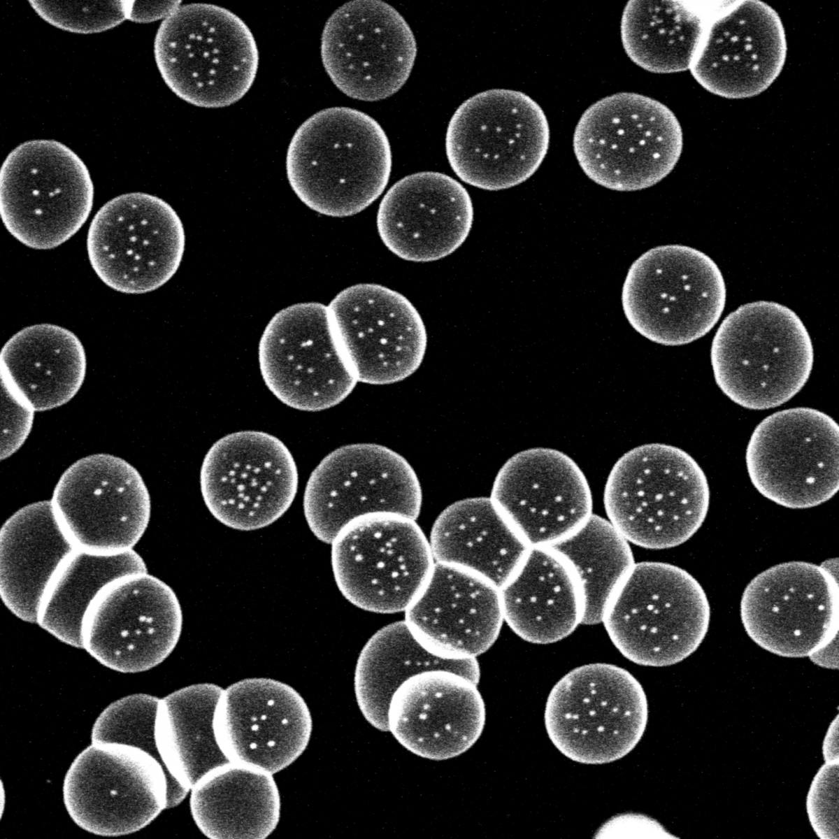
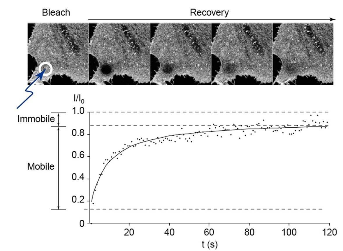
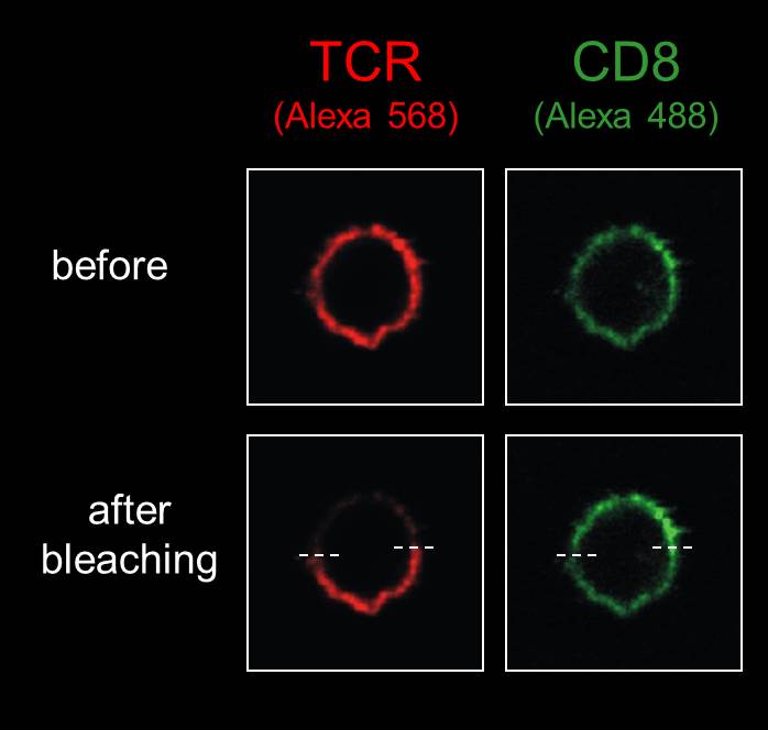
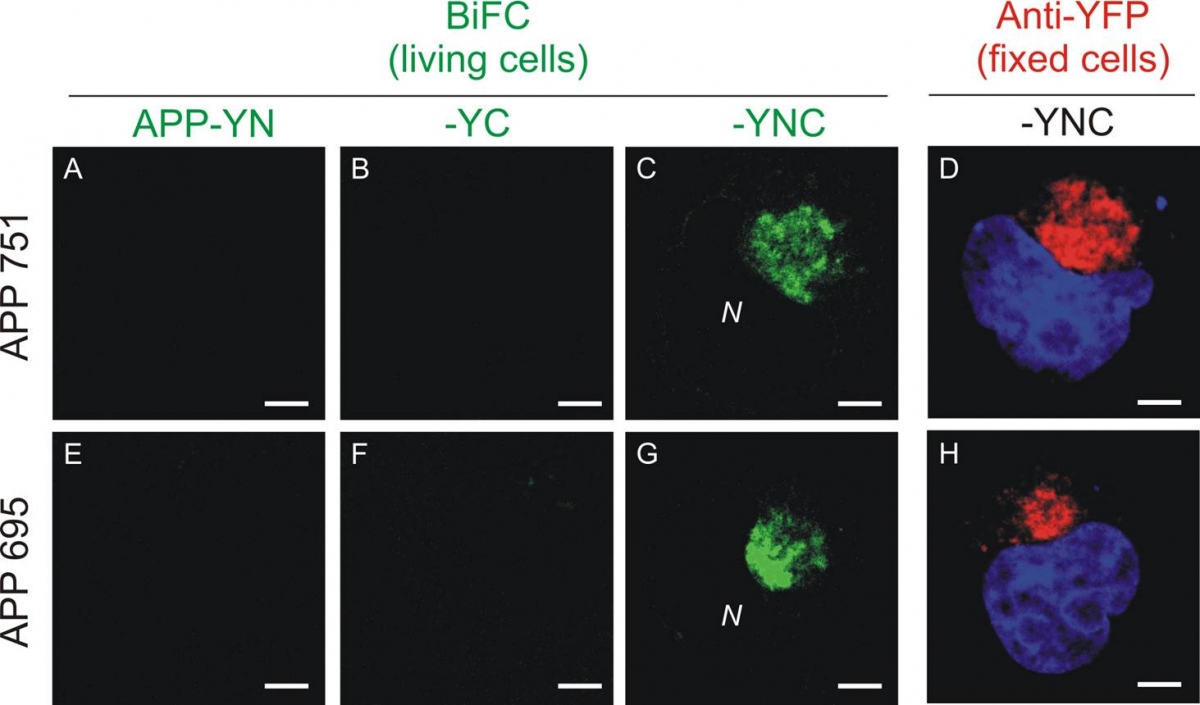
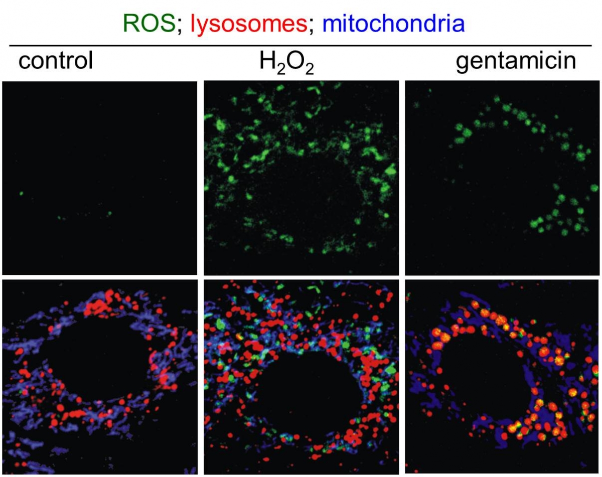
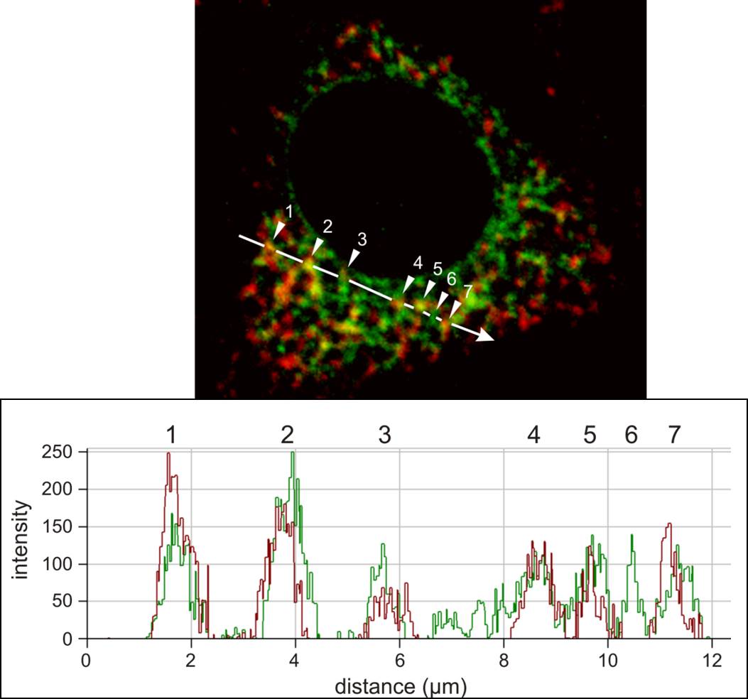
We are happy to share expertise in scientific projects addressing defined questions which can benefit from: (i) electron microscopy (scanning and transmission), including ultrastructural cytochemistry; (ii) high-resolution and high-throughput confocal microscopy, for live-cell imaging and immunolabelling; (iii) multiphoton microscopy to image tissues and organs for several hours without damage; and (iv) advanced applications by confocal microscopy, including dynamics of molecular movement by Fluorescence Recovery After Photobleaching (FRAP), protein dimerization by Bimolecular Fluorescence Complementation (BiFC), in situ detection of free radicals (ROS) at distinct subcellular compartments, and protein:protein interactions by FRET, etc. In addition, we propose advanced images analyses such as deconvolution, 3-D reconstruction, morphometry, objective co-localization, etc.
EXPERTISES
- Basic training and supervision of routine imaging |
- Sophisticated methods for preparation of biological samples (cells, tissues and organs) |
- Advanced training destined for independent, registered platform users |
- Vital cell and subcellular imaging with particle tracking |
- Fluorescence imaging on fixed cells and tissues.
|
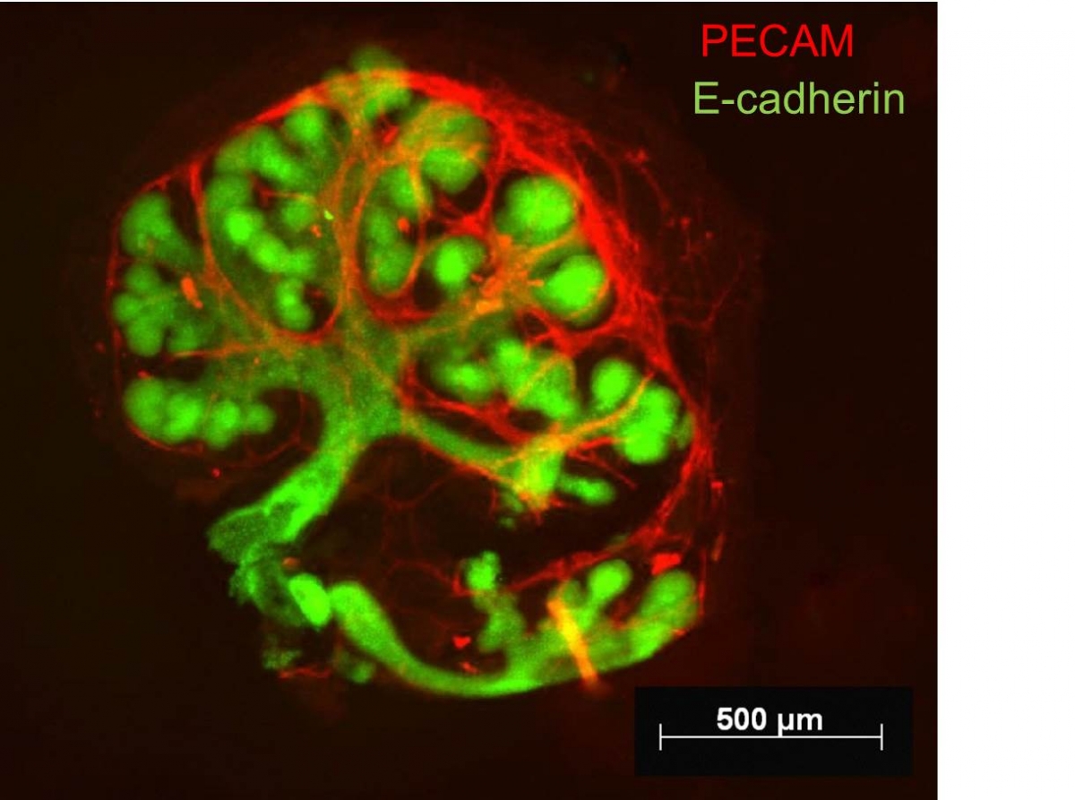
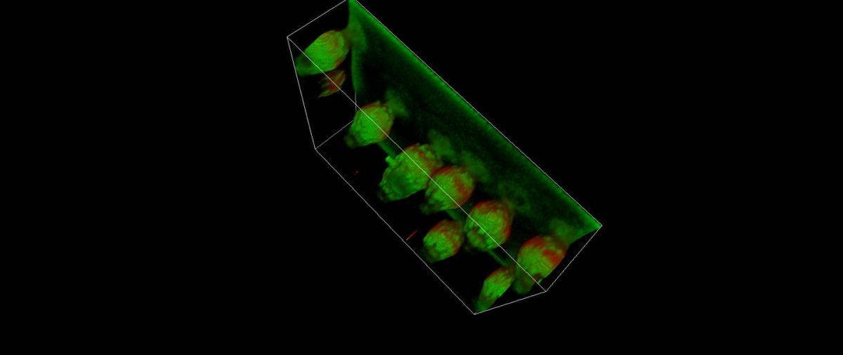
- Extended tissue and organ imaging without damage by multiphoton microscopy |
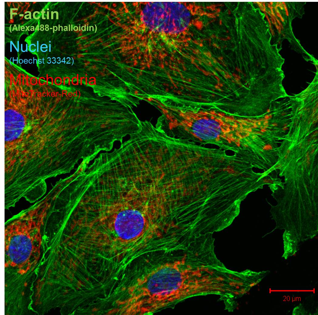
- High-resolution confocal imaging on fixed cells, tissues and organs.
|
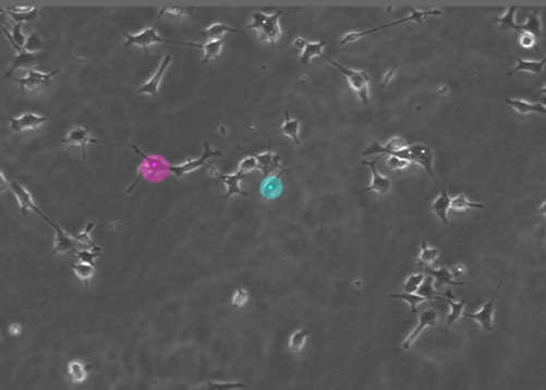
- Fluorescence imaging on living cells: cell migration.
|
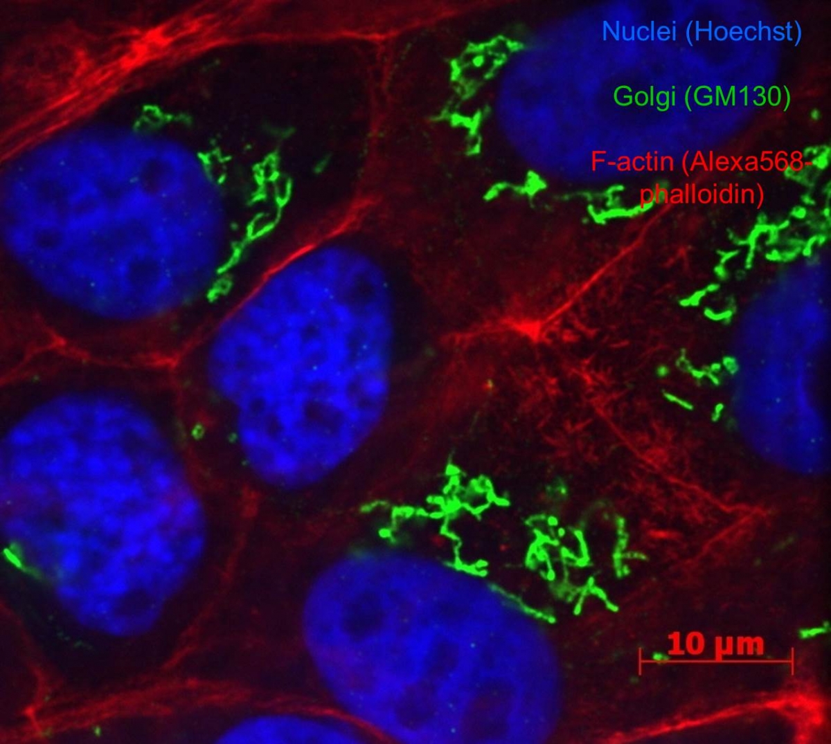
- High-throughput confocal microscopy.Triple (immune)labelling.
|
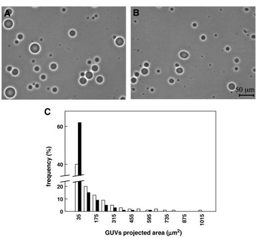
- High-resolution confocal imaging on living cells.Vital imaging of stable submicrometric membrane lipid domains (D’auria et al, 2013).
|
- Advanced applications by confocal microscopy:
|
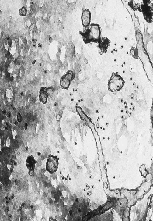
- Electron microscopy:
|
|
|
- Advanced off-line image analysis
|

