DIGITAL WHOLE SLIDE SCANNING MICROSCOPY
DESCRIPTION
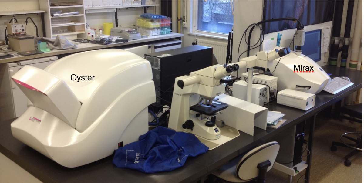
A first slide scanner (Zeiss Mirax Midi) was acquired in 2008. It comprises a microscope with a 20x objective coupled to a 1,4.106 px camera for both brightfield and fluorescence image acquisition, an automated 12-slide holder, a high-pressure mercury short-arc lamp and 5 filter sets for fluorescence scanning, and a computer.
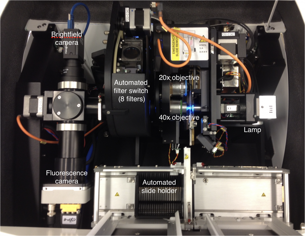
A second slide scanner (3DHistech Pannoramic P250 Flash III, nicknamed Oyster), with much improved scanning speed and slide capacity, and with better fluorescence performance, was acquired in 2017. It comprises a microscope with 2 objectives (20x and 40x), two interchangeable cameras (a 1,2x107 px high-frame colour camera for brightfield and a 4x106 px high-sensitivity monochrome camera for fluorescence image acquisition), a 250-slide automated holder, a light source with 5 LED lamps and 8 filter sets for fluorescence scanning, and a computer.
Both imaging platforms were acquired thanks to a grant from the Fonds de la Recherche Scientifique. They are accessible to all researchers from the UCLouvain-Woluwe campus, and to external users under conditions.
 |
|
Detailed view inside the Oyster slide scanner |
The slide digital imaging platform |
|
|
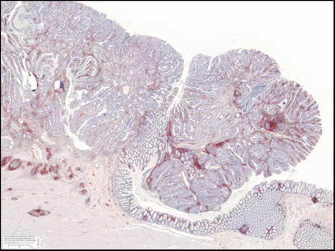
| Example image of a scanned microscopy image of a human colon tumor. Click on the image to open a new window that allows you to enlarge the view and navigate. Cell nuclei are stained in blue. A specific immunostaining for cytolytic CD8+ T lymphocytes (black) and helper CD4+ T lymphocytes (red) was applied, allowing to visualize the abundance and distribution of these immune cells in the tumor. |
APPLICATIONS
Digital acquisition of histology, immunohistology and immunofluorescence microscopy images.
As compared with conventional microscopy, digital microscopy has the following advantages:
- The publication of histological images is considerably improved. Traditionally, researchers publish microscope pictures from highly selected tissue areas and samples, which are very rarely representative of the entire histological sections and sample series. By publishing the entire digital images, we gain in objectivity.
- Automated quantitative data analysis on entire tissue sections is now possible thanks to the availability of powerful computers and efficient morphometry softwares such as ImageJ, SlidePath, and Visiopharm. For example, one can measure the total number of nucleated cells, the number and proportion of immunostained cells, the density of blood vessels, the surface of fibrotic tissue, etc. on large series of samples. This also contributes to the precision and objectivity of the data, and allows statistical analyses.
- The images acquired on multiple fluorescence channels can be superimposed digitally, allowing to study the co-expression of phenotypic markers in individual tissue sections.
- Image comparison, e.g. different immunostainings performed on adjacent sections, is greatly facilitated, as the images can be placed side by side on the same computer screen.
- Data sharing for second advices, teaching purposes and scientific collaboration is simplified, as the digital images can be exchanged via storage media, computer servers or via the internet.
- The storage and archiving of images is easier. Unlike archiving of histological slides, which are fragile and whose stainings fade with time, there is no data loss.
- Time saving by automated handling is significant compared to the many manipulations required by optical microscopy systems coupled to a digital camera.
COSTS & CONTACTS
- Cost:
Pannoramic Midi ('Mirax') slide scanner : brightfield scanning : 1,0 €/h / fluorescence scanning : 2,5 €/h
These costs cover maintenance and repair services and spare parts, and are subject to adjustments on a yearly basis.
Pannoramic P250 Flash III ('Oyster') slide scanner : brightfield scanning : 2,5 €/30min / fluorescence scanning : 2,5 €/h
These costs cover personnel in charge, maintenance and repair services and spare parts,
and are subject to adjustments on a yearly basis.
- Contact: Nicolas van Baren


